|
|
Lymphedema
General Considerations
- Abnormal collection of protein-laden fluid in the soft tissues from lymphatic obstruction
- This, in turn, leads to extravascular accumulation of water and soft tissue swelling
- Affects primarily lower extremities (80%)
- Primary form involves a congenital defect in lymphatic system and may be associated with Turner, Klinefelter, and Noonan Syndromes, trisomy 21, 13 or 18
- Secondary form may be due to neoplasm, filariasis, obesity, trauma or surgery/radiation therapy
Clinical Findings
- Chronic swelling of an extremity
 (not the same patient as radiograph) (not the same patient as radiograph)
- Fever, chills, weakness
- Redness and thickening of skin
- Impairment of activities due to size and weight of extremity
- Non-tender pitting edema progressing to non-pitting edema
- Elephantiasis nostra verrucosa primarily on the shins
Imaging Findings
- Diagnosis is made clinically
- Imaging is generally not needed
- MRI may show causes of obstruction
Differential Diagnosis
- Congestive heart failure
- Chronic venous stasis
- Deep vein thrombosis
- Filariasis
Complications
Treatment
- Pharmacotherapy includes benzopyrones, retinoid-like agents, topical skin products and anthelminthic agents
- Compression stockings and physical therapy
- Elevation of limb

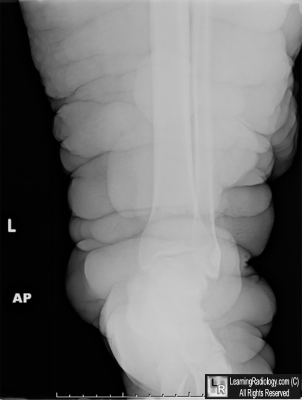
Lymphedema. There is marked soft tissue swelling of the left lower extremity. The patient had a negative workup for filarial disease and the leg was normal.
For this same photo, click here
For more information, click on the link if you see this icon 
Lymphedema. KM Rossy and NS Scheinfeld. eMedicine
|
|
|
